The Invisible Arsonist: How Fructose, Cholesterol, and Inflammation Together Destroy Our Arteries
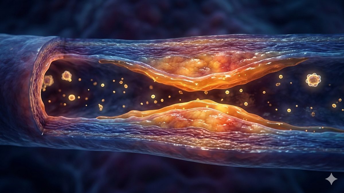
The article analyzes how fructose, cholesterol, and inflammation jointly contribute to the hardening of the arteries. Drawing on a range of studies, it explains that fructose not only raises the level of atherogenic lipoproteins in the blood, but at the same time damages the vascular wall through oxidative stress, a loss of nitric oxide, and chronic inflammation. The scientific consensus shows that atherosclerosis is not caused by any single factor, but by the convergence of the lipid and vascular axes, with fructose from added sugars acting as a powerful accelerator of the entire process.
In 2009, Kimber Stanhope of the University of California, Davis published a study that would change the way we think about heart and vascular disease. Thirty-two overweight volunteers drank sweetened beverages for ten weeks — one group's drinks sweetened with glucose, the other's with fructose. Both groups consumed the same number of calories. Both gained roughly the same amount of weight. But when the physicians drew blood, the results differed dramatically. The fructose group had higher LDL cholesterol, more small dense LDL particles, higher apolipoprotein B, higher postprandial (measured after eating) triglycerides, and lower insulin sensitivity. The glucose group — none of that.
The study, published in the Journal of Clinical Investigation, opened a question that medicine has not fully answered to this day: is the chief culprit behind the hardening of the arteries cholesterol, fructose, or something else entirely?
For fifty years, a simple equation dominated cardiology: high LDL cholesterol equals clogged arteries. Statins, low-fat diets, target LDL values — the entire prevention system rests on this premise. Over the past decade, however, evidence has been accumulating that the equation is incomplete. The CANTOS study (2017) demonstrated that blocking inflammation reduces the number of events threatening the heart and blood vessels even without any change in cholesterol. Metabolic research has revealed that fructose — the main component of added sugars — damages blood vessels in ways that standard blood-lipid testing does not detect at all.
For anyone trying to understand what actually causes the hardening of the arteries and how to prevent it, it is essential to know how these three elements — fructose, cholesterol, and inflammation — work together. In this case, simplified answers do harm.
To understand the current state of knowledge, we have to go back to the 1960s, when two competing hypotheses about the causes of heart disease were pitted against each other. The American physiologist Ancel Keys argued that the main culprit was saturated fat and cholesterol. The British physiologist John Yudkin maintained that the problem was sugar — above all sucrose and its fructose component.
In 2016, the researcher Cristin Kearns of the University of California, San Francisco, while studying archival documents, revealed that in 1965 the sugar-industry association the Sugar Research Foundation had paid two Harvard scientists the equivalent of roughly $48,000 (in 2016 dollars) for a review article in the New England Journal of Medicine that downplayed the role of sugar and directed attention to fats. Keys won. Yudkin was pushed to the margins. The result was four decades of dietary recommendations focused on reducing fat — during which the consumption of added sugars rose sharply.
Modern science vindicates both of them, but with nuances that neither could have suspected.
Fructose differs from glucose in one fundamental respect: its processing in the liver is not subject to feedback regulation. Glucose is processed by all the body's tissues, and the key enzyme in its breakdown — phosphofructokinase — halts once the cell has enough energy. At higher doses, fructose is largely captured by the liver on first pass and phosphorylated by the enzyme ketohexokinase, which has no such brake. (At low doses, most fructose is already processed by the small intestine — see below on the gut-liver axis.)
The result is predictable: an uncontrolled supply of carbon units into the fat-building pathways. The liver makes fat from fructose — a process called de novo lipogenesis (DNL). A randomized controlled trial by Geidl-Flueck et al. (2021, Journal of Hepatology, n = 94 healthy lean men) quantified it: 80 grams of fructose per day, after seven weeks, doubled baseline hepatic DNL. The same dose of glucose had no measurable effect.
The newly created fats are packaged into VLDL particles and exported into the blood. This is where a chain reaction begins that alters the entire lipid profile. The study by Stanhope et al. (2015, American Journal of Clinical Nutrition, n = 85) showed a direct dose-response relationship between high-fructose corn syrup and the rise in LDL cholesterol: at 25% of energy intake, LDL-C increased by 15.9 mg/dL above baseline (in the control group with no sweetener, it did not change).
But the LDL-C number itself is only the tip of the iceberg. More important is what happens to the properties of the LDL particles. Elevated triglycerides in VLDL trigger a chain reaction: the CETP protein exchanges triglycerides from VLDL for cholesteryl esters from LDL — producing a triglyceride-enriched LDL — hepatic lipase cleaves it — and the result is small dense LDL particles (sdLDL). These particles are highly atherogenic — they penetrate the vessel wall more easily, circulate in the blood longer (the LDL receptor recognizes them less readily), and are more prone to oxidation.
A review analysis by Zhang et al. (2013, Journal of Nutrition, 24 studies) confirmed an increase in LDL-C, but only at doses above 100 g of fructose per day. By contrast, a review analysis by Chiavaroli et al. (2015, Journal of the American Heart Association, 59 studies) found that under isocaloric substitution — that is, when fructose replaces other carbohydrates without increasing total calories — LDL-C, triglycerides, and HDL-C do not change significantly. The key insight: fructose harms the lipid profile chiefly when it represents a caloric excess. And in the practice of the modern diet, it almost always does.
Now comes the question that is crucial to understanding the whole problem: if fructose alters the blood lipid profile, is it cholesterol that damages the arteries? Or are there other pathways as well?
The consensus statement of the European Atherosclerosis Society (Ference et al., 2017, European Heart Journal) answers the first question unequivocally: lipoproteins carrying apolipoprotein B — that is, LDL, VLDL remnants, and lipoprotein(a) — cause atherosclerotic cardiovascular disease. The evidence base includes Mendelian randomization studies, large epidemiological cohorts, and over 200 randomized clinical trials of LDL-lowering drugs.
The key model of the onset of atherosclerosis was described by Williams and Tabas (1995, Arteriosclerosis, Thrombosis, and Vascular Biology) as the "response-to-retention." What is decisive is not the penetration of LDL into the vessel wall itself — that occurs in all people. What is decisive is its retention in the subendothelial space through binding to proteoglycans. The retained LDL gradually oxidizes, is engulfed by macrophages via scavenger receptors, the macrophages transform into foam cells, and from these the atherosclerotic plaque arises.
But — and this "but" changes the whole picture — cholesterol is not the only player. In 2017, the CANTOS study (Ridker et al., New England Journal of Medicine, n = 10,061) delivered groundbreaking evidence: canakinumab, an antibody against interleukin-1β, reduced cardiovascular risk by 15% (hazard ratio 0.85) without changing LDL-C, HDL-C, or triglycerides in any way. It worked purely by reducing inflammation. The follow-up studies COLCOT (2019) and LoDoCo2 (2020) with colchicine confirmed the results.
Peter Libby of Harvard Medical School summed it up in a landmark review article (2021, Nature): inflammatory pathways link both traditional and new risk factors to the altered behavior of vessel-wall cells. Atherosclerosis is a disease driven by lipids, amplified by inflammation, and influenced by many factors at once.
And this is exactly where fructose enters the game as something far more dangerous than a mere supplier of cholesterol.
Picture atherosclerosis as a fire. Cholesterol (more precisely, apoB-containing lipoproteins) is the combustible material — without it, there is no fire. But fructose is the arsonist, who simultaneously adds more combustible material and pours gasoline over the surroundings.
Proof that the combustible material remains essential came from animal models. Merat et al. (1999) demonstrated in LDL-receptor-deficient mice that, at comparable blood cholesterol levels, a fructose diet did not induce more atherosclerosis than the control — even though it induced insulin resistance. Similarly, Kostogrys et al. (2012, Atherosclerosis) found in apoE- and LDL-receptor-deficient mice that a fructose diet without an increase in cholesterol did not enlarge the atherosclerotic lesions. Without apoB lipoproteins in the vessel wall, plaque simply does not form.
But that is only half the story. Fructose damages the vascular system in at least five ways that are formally independent of classic cholesterol.
Advanced glycation end products of non-enzymatic glycation. Fructose is eight to ten times more reactive than glucose in the Maillard reaction — the non-enzymatic joining of sugars to proteins. The reason is chemical: fructose has a higher proportion of the open-chain form, which is reactive. The resulting products — fructose-AGEs — activate the RAGE receptor on endothelial cells, induce the formation of the adhesion molecule VCAM-1, and generate reactive oxygen species (Sotokawauchi et al., 2019, Diabetes & Vascular Disease Research). The fundamental problem: routine clinical tests such as HbA1c or fructosamine do not detect these products, because they measure only glucose glycation.
Oxidative stress. Delbosc et al. (2005, Atherosclerosis) demonstrated in rats on a fructose diet an increased production of reactive oxygen species via NADPH oxidase as early as one week in — before blood pressure had even risen. Carvalho Braga et al. (2015) confirmed the direct activation of NADPH oxidase by fructose in the isolated aorta.
Nitric oxide depletion. Nitric oxide (NO) is a key vasodilator and a protective factor of the inner lining. Stirpe et al. (2022) found that even physiologically relevant concentrations of fructose (0.1–1 mM), under intermittent exposure, caused an 80–90% drop in NO production in endothelial cells — and the effect persisted for 12 hours after the fructose was removed. An endothelium without NO is more permeable to LDL, more prone to inflammation, and more prone to the formation of blood clots.
Uric acid and xanthine oxidase. The phosphorylation of fructose by ketohexokinase consumes ATP without regulation. The accumulating AMP is broken down via xanthine oxidase into uric acid — and in the process, xanthine oxidase generates superoxide. Baldus et al. demonstrated that inhibiting xanthine oxidase improves the function of the vessel wall lining, whereas merely lowering uric acid with drugs that promote its excretion does not. The harmful agents are therefore primarily the radicals from the enzymatic reaction, not just the end product.
Direct induction of inflammation. Shen et al. (2025, FASEB Journal) described how fructose activates macrophages via the ghrelin receptor GHSR and increases the pro-inflammatory cytokines IL-1β, IL-6, and TNF-α. Cirillo et al. (2015) demonstrated that fructose induces the production of tissue factor in endothelial cells — creating an environment conducive to clot formation.
The two axes amplify each other. Fructose produces more atherogenic lipoproteins (sdLDL, VLDL remnants, apoB) and at the same time prepares an environment of oxidative stress, inflammation, and a damaged endothelium that facilitates their retention and oxidation in the vessel wall.
It would be convenient to end with a simple verdict: fructose is the poison, cholesterol is the accomplice, end of story. But science does not work so simply, and there are serious counterarguments.
First, under isocaloric conditions — when fructose replaces other carbohydrates without an increase in total calories — most of the adverse effects on the lipid profile are markedly weakened or disappear. This is a consistent finding of the review analyses by Chiavaroli et al. (2015) and others. One can argue that the problem is not fructose as such, but the caloric excess that the consumption of sweetened beverages facilitates.
Second, whole fruit — which contains fructose — demonstrably protects against cardiovascular disease. A review analysis by Sun et al. (2023, Advances in Nutrition, 64 long-term follow-up studies) quantified it: each serving of fruit per day reduces the risk of cardiovascular disease by 3–6%, whereas every 250 ml of sweetened beverages increases it by 10%. The fiber in fruit slows the absorption of fructose, lowers the dose reaching the liver, and provides anti-inflammatory plant compounds. The context and the form of the source are decisive.
Third, the conflicts of interest on both sides of the dispute must be mentioned. Some of the review analyses that downplay the harmfulness of fructose were funded by food-industry organizations (the Calorie Control Council, the Corn Refiners Association, the International Sweeteners Association). But even some researchers on the "anti-fructose" side — such as Robert Lustig — are criticized for excessive oversimplification. The scientific truth lies somewhere in the middle.
The current scientific picture can be summed up by a model of two converging axes.
The lipid axis: Fructose increases hepatic de novo lipogenesis → more VLDL → more triglycerides → conversion of LDL into small dense particles → higher apoB → more atherogenic lipoproteins penetrating the vessel wall.
The vascular axis: Fructose produces advanced glycation products, activates NADPH oxidase, depletes NO, raises uric acid and the radicals generated by xanthine oxidase, activates macrophages → damage to the vessel-wall lining, oxidative stress, chronic inflammation → easier retention and oxidation of lipoproteins in the vessel wall.
Without apoB lipoproteins, plaque does not form — this is shown by animal models with knocked-out genes. But with fructose it forms faster, from a smaller quantity of lipoproteins, and in a more aggressive form.
A review analysis by Khan et al. (2019, Mayo Clinic Proceedings, 624,128 people) identified a threshold dose for death from cardiovascular disease: 58 g of fructose per day, which corresponds to roughly 11% of energy intake. Below this threshold no significant association was found. It must be added, however, that the authors themselves rated the quality of the evidence base under the GRADE system as "low," and the study was partly funded by the industry association the Calorie Control Council. Yang et al. (2014, JAMA Internal Medicine) found that people whose added-sugar intake exceeded 25% of calories had nearly three times the risk of death from cardiovascular disease compared with those below 10%.
The biological basis for the existence of a threshold dose is provided by the discovery of the gut-liver axis: low doses of fructose are processed mainly in the small intestine by the enzyme ketohexokinase-A. Only when the intestine's capacity is exceeded does fructose "spill over" into the liver, where it triggers de novo lipogenesis and other pathological processes.
The current recommendations of the World Health Organization, the American Heart Association, and the European Food Safety Authority target added sugars in general: the WHO recommends less than 10% of energy from free sugars (ideally below 5%, that is, roughly 25 g per day), while the AHA has set a ceiling of 25 g per day for women and 36 g for men. No major recommendation distinguishes between fructose and other added sugars — which, on the one hand, simplifies the message, but on the other hand obscures the fact that fructose has unique metabolic properties.
Thirty-two volunteers in Davis, California. Two groups. The same calories. The same weight gain. But a fundamentally different lipid profile, fundamentally different visceral fat stores, fundamentally different insulin sensitivity. Fructose was not just an "empty calorie." It was a metabolic program that rewrote the biochemistry of the liver, the blood, and the vessel wall — and standard testing largely overlooked this rewriting.
Seventeen years later, we know that the hardening of the arteries is caused neither by "cholesterol alone," nor by "fructose alone," nor by "inflammation alone." It is the convergence of all three — and within it, fructose functions as a powerful accelerant, which increases the quantity of the most dangerous lipoproteins while at the same time weakening the defenses of the very blood vessels that are supposed to resist them.
John Yudkin did not have all the details right. But his fundamental insight — that sugar is more dangerous to the heart than fat — is, after fifty years, proving to be closer to the truth than the scientific mainstream ever admitted.
This article is based on studies published in peer-reviewed professional journals (JCI, NEJM, European Heart Journal, JAHA, AJCN, Journal of Hepatology, Nature, Mayo Clinic Proceedings, JAMA Internal Medicine, Frontiers in Nutrition, Advances in Nutrition). The principal sources are randomized controlled trials, review analyses, and consensus statements of professional societies.
Main limitations: most intervention studies with fructose lasted 2–10 weeks, which does not allow long-term effects to be assessed. The doses in randomized trials often exceed ordinary consumption (25% of energy intake). Animal models with knocked-out genes have limited transferability to humans. Long-term follow-up studies cannot prove a causal relationship. A number of studies on both sides of the dispute have acknowledged or possible conflicts of interest with the food industry.
Open questions: the precise threshold dose for different population groups, the role of intestinal processing as a protective filter, the causal relevance of fructose-specific mechanisms of action (the ghrelin receptor, cardiac ketohexokinase-C) in humans, and the interaction of fructose with other components of the modern diet (industrially processed foods, saturated fats, salt).
Transparency of creation
The concept, structure, and editorial line of the article are the work of the author, who prepared the content outline, established the key theses, and directed the entire creative process. Generative AI (Claude Opus 4.6, Anthropic) was used as a tool for research, fact-checking, and fleshing out the author's draft.
The author verified the key findings and approved the final wording. No part of the text was published without conscious authorial oversight. The factual data were verified against the publicly available sources cited in the text.
The procedure complies with the transparency principles of EU Regulation 2024/1689 (the AI Act). #poweredByAI
Read the Czech original on Médium.cz.
AI · Claude — machine translation, may contain inaccuracies.