The Korean Paradox: Why Pork Belly Doesn't Kill You, but Cola Does
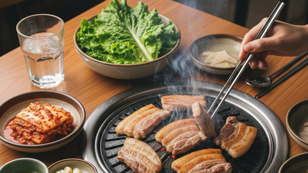
The article explains why Koreans, despite their high consumption of fatty pork belly, have significantly lower circulatory mortality and obesity than Czechs. The key is their low intake of fructose and sweetened drinks — new studies from 2024–2025 have revealed that fructose causes fatty liver via two independent mechanisms (classic de novo lipogenesis and the newly discovered follistatin pathway), with the combination of high fat and high fructose being the most metabolically dangerous.
In Seoul, South Korea, thousands of restaurants specializing in samgyeopsal — slices of pork belly grilled right at the table — light up every evening. Three layers of fat, no compromises. Koreans eat 30 kg of pork per person each year, and belly is by far the most popular cut — 60% of respondents in a survey by the Korea Rural Economic Institute (KREI) named it their first choice. Fat content: around 30%, of which almost half is monounsaturated fatty acids (mainly oleic acid), roughly a third saturated, and the rest polyunsaturated.
According to the logic that dominated nutritional guidelines for half a century — saturated fats clog arteries and cause heart disease — Korea ought to be a cardiovascular catastrophe. Yet its age-standardized mortality from cardiovascular disease runs at roughly 65–90 deaths per 100,000 inhabitants, depending on the methodology and reference population. In the Czech Republic, where total pork consumption is even higher (43 kg per year) but belly is not the dominant cut, cardiovascular mortality reaches approximately 270–330 per 100,000 (men markedly more than women). Obesity? Korea 5.9% of adults with a body mass index above 30, the Czech Republic 26%. Life expectancy? Korea 83.7 years, the Czech Republic 80 years.
Something doesn't add up. And a series of studies from 2024–2025 finally explains what.
In February 2026, Medium.cz published the article "How the sugar industry stole half a century of nutritional science," which traced how the Sugar Research Foundation (SRF) paid Harvard scientists in 1965 for a review article that then appeared in 1967 in the New England Journal of Medicine. The article successfully redirected attention from sugar to fat as the principal culprit behind cardiovascular disease. John Yudkin, the British professor of nutrition at Queen Elizabeth College, University of London, who had warned against sugar, was pushed to the margins. Ancel Keys, with his hypothesis about saturated fats, won.
This article picks up where the previous one left off. Yudkin was right — but even he had no idea exactly how sugar does its damage. New studies from 2025 reveal a mechanism substantially more complex and more dangerous than Yudkin could have imagined. Fructose does not merely create fat directly in the liver. As a groundbreaking study in Nature Communications from November 2025 shows, fructose also causes fatty liver by a route we previously knew nothing about.
Until now, the consensus held that fatty liver (MASLD — metabolic dysfunction-associated steatotic liver disease, formerly known as NAFLD) requires insulin-stimulated synthesis of new fats (de novo lipogenesis). Simplified: insulin activates the transcription factor SREBP-1c in the liver, which switches on the enzymes for building new fats from surplus carbohydrates, and these fats are deposited in liver cells. Without the insulin signal — no fatty liver. That was the model.
In November 2025, the team of Morris F. White from Boston Children's Hospital (Harvard Medical School) published a study in Nature Communications that shatters this model. The researchers worked with mice in which both hepatic insulin receptor substrates had been genetically knocked out (so-called LDKO mice) — thereby creating a state of complete hepatic insulin resistance. On a high-fat diet, these mice never developed fatty liver, exactly as the prevailing model predicted.
But when fructose was added to these mice's diet, they developed acute MASLD. And this despite the fact that the insulin signaling pathway in their livers did not function at all.
How is that possible? The team demonstrated that in LDKO mice, fructose did not act through classical de novo lipogenesis — no new fatty acids were synthesized. Instead, fructose provided a glycerol backbone for the re-esterification of free fatty acids flowing into the liver from adipose tissue. Carbon-13 isotope labeling showed that after labeled fructose was administered, ¹³C was incorporated into the glycerol backbone of triacylglycerols, but not into their fatty acids. The fat therefore did not come from new synthesis — it came from the body's fat stores, which fructose helped to "repackage" and deposit in the liver.
Follistatin (FST) — a hormone produced by the liver — plays a key role in this process. In LDKO mice on a fructose diet, follistatin production was markedly increased. Follistatin blocks activin signaling in adipose tissue, thereby increasing the insulin resistance of fat cells — adipose tissue then massively releases fatty acids into the blood. These acids accumulate in the liver, where fructose re-esterifies them. When the researchers genetically inactivated hepatic follistatin in LDKO mice, acute MASLD did not develop. Conversely, when they overactivated follistatin in healthy mice, fatty liver accelerated dramatically.
Human data confirmed this: in participants of the Tübingen Diabetes Family Study (TDFS), higher blood follistatin levels were associated with greater insulin resistance of fat cells and higher triacylglycerol content in the liver.
The implication is fundamental: fructose causes fatty liver via two independent routes — classical de novo lipogenesis through SREBP-1c (when insulin signaling works) and re-esterification through the follistatin pathway (even when insulin signaling does not work). It is a double lock, not a single mechanism.
In June 2025, another piece was added to the puzzle by a study from Parama Bhattacharjee and colleagues at the University of Maryland, published in the Journal of Nutrition. The researchers fed mice a combination of a high-fat diet with fructose for 24 weeks and observed what was happening in the liver mitochondria.
The result was unexpected and disturbing. Fructose simultaneously induced de novo lipogenesis and mitochondrial oxidative function — that is, both the creation of fats and their burning. This sounds contradictory, but it is precisely this "dual induction" that is metabolically devastating. The mitochondria, forced to burn ever more fats, produce increased amounts of reactive oxygen species (ROS). These free radicals damage cellular structures, trigger inflammation, and, according to the study's authors, could accelerate the transition from simple steatosis (MASLD) to the more dangerous steatohepatitis (MASH) with a risk of fibrotic remodeling.
The authors conclude that fructose creates a state of chronic metabolic strain in the liver — the mitochondria run at full power, but at the same time fats accumulate that they cannot keep up with processing. It is as if you were pressing the accelerator and the brake at the same time.
The Brazilian team of Flávia Maria Silva-Veiga from the State University of Rio de Janeiro published a study in March–April 2025 in Obesity Research & Clinical Practice directly comparing four groups of mice: control diet, high-fat diet, high-fructose diet, and the combination of both. All three intervention groups developed hepatic steatosis, mitochondrial disturbances, and endoplasmic reticulum stress, but the worst damage was shown precisely by the combination of high fat with high fructose — increased production of proteins associated with inflammation, lipogenesis, and cellular stress.
At the same time, in October 2025, a Chinese team (Yu Z, Chen M, Gu S et al.) published a study in Frontiers in Endocrinology of 3,305 analyzed patients with type 2 diabetes, in which sugar-sweetened beverage consumption predicted fatty liver independently of other risk factors. And a large British study by Zhou Y and colleagues in Frontiers in Public Health (2025) analyzed magnetic resonance images of the liver in 25,885 UK Biobank participants — both sugar-sweetened beverages and artificial sweeteners were associated with higher liver fat content and inflammation of liver tissue.
A systematic review by Yu ZZ and colleagues (2025), published in Frontiers in Nutrition and prepared for presentation at the conference of the American Association for the Study of Liver Diseases (AASLD), summarized the evidence on high-fructose corn syrup (HFCS) and liver injury: controlled studies show that HFCS in beverages has negative effects on liver fat content and insulin sensitivity, although the evidence base (only one study out of 23,006 assessed met the strict criteria) does not yet allow a definitive conclusion about a dose-dependent relationship.
These findings change the view of the Korean diet in a fundamental way. Koreans eat enormous amounts of fatty pork belly — but they consume it in a metabolic context that dramatically reduces its harmfulness.
Total sugar (sucrose) consumption in Korea is roughly 27–28 kg per person per year, compared with the Czech 37 kg. On top of that, Korea consumes about 5–6 kg of high-fructose corn syrup per person per year — a fraction of American consumption. Sugar-sweetened beverage consumption in Korea is indeed rising, but the baseline level remains low compared with the West.
The key, however, is not just what Koreans don't drink, but what they eat in addition. The traditional Korean diet stands out for its high proportion of vegetables — Korea ranks among the leaders within the OECD countries. Kimchi, fermented vegetables consumed by a large majority of the adult population daily in portions of around 50–200 g (albeit with a declining trend), provides probiotics (bacteria of the genus Lactobacillus), fiber, and bioactive compounds. Seafood consumption reaches 61 kg per year — one of the highest figures in the world — and supplies omega-3 fatty acids with a proven protective effect on the cardiovascular system. Fiber intake is 23.2 g per 2,000 kcal per day, well above European values.
Samgyeopsal, moreover, is typically eaten wrapped in fresh lettuce (ssam) with garlic, kimchi, and fermented soybean paste — not with white-flour dumplings and beer.
For comparison: Czech dietary patterns combine saturated fats with refined carbohydrates (dumplings, white bread), high beer consumption (around 128–152 liters per year depending on methodology — long-term first place in the world), and relatively low intake of vegetables and fish. Precisely the combination of high fat with high fructose and refined carbohydrates that the 2025 studies identify as the most metabolically dangerous.
It would be tempting to declare that Korea has it solved and the Czech Republic does not. The reality is more nuanced.
First, the genetic predispositions differ. The Korean population has a different frequency of gene variants associated with fat and alcohol metabolism — including PNPLA3 (the risk allele occurs in ~45% of Koreans compared with ~23% of Europeans) and ALDH2 (a mutation typical of East Asia, practically absent in Europeans). These genetic differences may influence the metabolic response to saturated fats independently of diet. A population comparison is not a controlled experiment.
Second, the Korean metabolic "bonus" is dissolving. The prevalence of metabolic syndrome in Korea rose from 27.1% in 2001 to 33.2% in 2020, and among men even to 36.8% (2022). Fat intake among Korean adolescents is climbing — the proportion of saturated fat rose from 7.1% to 8.4% of energy over 2007–2017. Beverage consumption more than quadrupled since 1998. Kimchi consumption is declining. The Westernization of the Korean diet is in full swing, and the metabolic consequences will come.
Third, Czech cardiovascular mortality is falling dramatically. From values exceeding 600 per 100,000 in the 1980s to the current roughly 270–330. The progress is real, even if still far from the Korean figures.
Fourth, factors outside diet also play a role — the level of the healthcare system, the degree of physical activity, smoking, stress, social cohesion. Korea has a very well-organized system of preventive check-ups and a markedly lower proportion of female smokers (around 4–6% compared with 16–22% in the Czech Republic).
And finally, it is necessary to distinguish studies on animal models from population data. The 2025 mouse studies provide mechanistic insight, but transferring it to the human population requires caution. Human fructose metabolism involves a first pass through the intestinal wall, where at low doses a significant portion of fructose is converted into glucose and organic acids — something the mouse models with high doses do not fully capture.
Despite these reservations, the convergence of evidence from mechanistic studies, controlled human trials (Geidl-Flueck et al. 2021: 94 men, randomized controlled trial), and population data is consistent. Fructose is a metabolic amplifier — it amplifies the damage from fat, and an environment poor in fructose (the Korean model) markedly attenuates that damage.
For readers who want to understand the mechanism more deeply, a brief summary of how fructose works in the liver — and why it is different from glucose.
When you eat glucose, it enters glycolysis, where the key enzyme phosphofructokinase acts as a "brake" — when energy is abundant, glycolysis slows down. Glucose is also processed in the muscles, brain, and other tissues. The liver processes roughly a quarter to a third of incoming glucose.
Fructose is different. Almost all fructose heads straight to the liver, where the enzyme fructokinase C (KHK-C) immediately phosphorylates it — without any feedback regulation. Fructose bypasses phosphofructokinase, the main regulatory point of glycolysis. It is fuel that pours into the metabolic engine without brakes.
The consequences are threefold. First, rapid phosphorylation consumes ATP and, via the breakdown of purines, generates uric acid, which triggers oxidative stress and inflammation. Second, the surplus intermediates directly activate the transcription factor ChREBP and indirectly (via insulin signaling) also SREBP-1c — both switch on the enzymes of de novo lipogenesis, and the liver begins to manufacture fat massively. Third — and this is the new insight from 2025 — fructose stimulates the production of follistatin, which, through the insulin resistance of fat cells, releases fatty acids from adipose tissue and provides a glycerol backbone for their re-esterification in the liver.
The result: the liver fills with fat from three sources at once — from new synthesis, from the repackaging of fats from adipose tissue, and from impaired burning due to mitochondrial strain.
In Seoul, friends gather around a grill with pork belly. On the table are bowls of kimchi, fresh lettuce, garlic, spices, fermented paste. Water, green tea, or soju — but not a liter of cola.
At the other end of Eurasia, a Czech orders roast pork with dumplings and sauerkraut — saturated fat combined with white flour and sweet beer. Or fried cheese with fries and a cola. Precisely the combination that the 2025 studies identify as the most metabolically dangerous: high fat together with high fructose, a joint assault on the liver by two independent routes.
The Korean paradox is no paradox. It is proof that fifty years after the Sugar Research Foundation paid Harvard scientists to divert attention from sugar, science is finally returning to the right question. It is not about how much fat you eat. It is about how much sugar you drink.
Methodological note: The article draws on peer-reviewed studies published between 2021 and 2025, population data from the WHO, OECD, FAO, and national statistical offices (ČSÚ, KNHANES). Animal studies provide mechanistic insight, but their direct transfer to the human population requires caution. The Korea–Czech Republic population comparison is observational and cannot prove a causal relationship.
New research 2024–2025 (main studies):
Tao R, Stöhr O, Tok O et al. "Fructose and follistatin potentiate acute MASLD during complete hepatic insulin resistance." Nature Communications 16, 11595 (2025). doi:10.1038/s41467-025-66296-5
Bhattacharjee P, Fadlaoui A, Ryan CE et al. "Induction of Fructose Mediated De Novo Lipogenesis Coexists with the Upregulation of Mitochondrial Oxidative Function in Mice Livers." Journal of Nutrition 155(6):1768–1781 (2025). doi:10.1016/j.tjnut.2025.04.030
Silva-Veiga FM, Miranda CS, Santana-Oliveira DA et al. "Excessive dietary fat and fructose enhance hepatic lipogenesis and impair mitochondrial dynamics to cause MASLD in C57BL/6 mice." Obesity Research & Clinical Practice 19(2):138–145 (2025). doi:10.1016/j.orcp.2025.03.007
Yu ZZ, Varahala S, Lim A, Marenah M, Wattacheril J. "The impact of high fructose corn syrup on liver injury and glucose metabolism: a systematic review." Frontiers in Nutrition 12:1724398 (2025). PMC12689413
Zhou Y, Zeng, Wan, Zhao et al. "Association of sugary beverages consumption with liver fat content and fibro-inflammation: a large cohort study." Frontiers in Public Health 13:1624848 (2025).
Yu Z, Chen M, Gu S et al. "Sugar-sweetened beverage consumption predicts metabolic associated fatty liver disease in patients with type 2 diabetes mellitus." Frontiers in Endocrinology 16:1651370 (2025).
Key earlier findings (context):
Geidl-Flueck B, Hochuli M, Niedermeyer A et al. "Fructose- and sucrose- but not glucose-sweetened beverages promote hepatic de novo lipogenesis: A randomized controlled trial." Journal of Hepatology 75(1):46–54 (2021). — 94 men, randomized controlled trial, 7 weeks.
Stanhope KL, Schwarz JM, Keim NL et al. "Consuming fructose-sweetened, not glucose-sweetened, beverages increases visceral adiposity and lipids and decreases insulin sensitivity in overweight/obese humans." Journal of Clinical Investigation 119(5):1322–1334 (2009).
Geidl-Flueck B, Gerber PA. "Fructose drives de novo lipogenesis affecting metabolic health." Journal of Endocrinology 257(2):e220270 (2023). PMC10083579 — review article.
Kearns CE, Schmidt LA, Glantz SA. "Sugar Industry and Coronary Heart Disease Research: A Historical Analysis of Internal Industry Documents." JAMA Internal Medicine 176(11):1680–1685 (2016). — archival discovery of SRF→Harvard funding.
Korean data:
Choe JH, Choi MH, Rha YA et al. "Characteristics of pork belly consumption in South Korea and their health implication." Journal of Animal Science and Technology 57:22 (2015). PMC4540268
Park D, Lim S et al. "20-Year Trends in Metabolic Syndrome Among Korean Adults From 2001 to 2020." JACC: Asia 3(3):491–502 (2023). PMC10308107
Korea Herald: "South Koreans consumed 30kg of pork per person in 2024." (2025).
Song S, Shim JE. "Trends in Dietary Intake of Total Fat and Fatty Acids Among Korean Adolescents from 2007 to 2017." Nutrients 11(12):3073 (2019). PMC6950604
Czech and comparative data:
OECD Health at a Glance 2023 / 2025: Country profiles — Korea, Czech Republic.
ČSÚ: Consumption of food and non-alcoholic beverages 2022–2024.
WHO Global Health Observatory: Age-standardized CVD mortality rates.
Cífková R et al. "30-year trends in major cardiovascular risk factors in the Czech population." BMC Public Health 20:585 (2020). PMC7213700
Transparency of creation
The concept, structure, and editorial line of the article are the work of the author, who prepared the content outline, established the key theses, and directed the entire creative process. Generative AI (Claude Opus 4.6, Anthropic) was used as a tool for research, fact-checking, and elaborating the author's outline.
The author verified the key findings and approved the final wording. No part of the text was published without conscious authorial oversight. The factual data were verified against the publicly available sources cited in the text.
The procedure complies with the transparency principles of EU Regulation 2024/1689 (the AI Act). #poweredByAI
Read the Czech original on Médium.cz.
AI · Claude — machine translation, may contain inaccuracies.